Two minutes into a consult, a runner pulls a crumpled MRI report from her bag and asks if she needs surgery for a “split peroneal tendon.” The scan looks dramatic. Her exam says something different. That gap between pictures and reality is exactly why preparation for imaging and tests matters when you meet a foot and ankle surgical evaluation doctor.
What your surgeon is actually trying to answer
A foot and ankle surgery doctor is not just hunting for a tear or a spur. The goal is to understand load, alignment, tissue quality, nerve status, and the pattern of your pain in real function. Imaging gives structure, tests give physiology, but the exam ties them to your symptoms. A foot and ankle surgical expert wants to know:
- Where the pain begins and where it travels when you walk, climb stairs, pivot, or push off. Whether the problem is primary bone, joint, tendon, ligament, or nerve, or a combination. If mechanics are the driver, such as flatfoot collapse, cavus alignment, forefoot varus, or equinus. Whether the pathology is acute, chronic, or an acute flare on chronic changes. What conservative care you have already tried, how strictly you followed it, and how your body responded.
The answers shape which tests matter and how the foot and ankle surgical provider interprets them. An MRI that looks ominous after a hard trail race can reflect normal changes in a stiff, strong tendon. A subtle instability that cripples a dancer can hide on routine ankle X‑rays unless stress views are done. Preparation, both yours and the clinic’s, reduces noise and exposes what truly needs attention.
Arriving ready: history that changes testing
The best imaging is built on a precise history. I ask for timelines in weeks and months, not “a while.” I listen for the first bad step, the voice of swelling, and what shoes you were wearing. Your preparation can save one or two follow‑up visits by clarifying simple details in advance.
Bring a simple pain map: point to one to three finger‑sized spots. Note when pain spikes, using activities rather than vague adjectives. “Pain 7 out of 10 after 20 minutes of walking on uneven ground” beats “it hurts a lot.” If morning pain eases after a few minutes, that suggests plantar fascia or Achilles involvement. If night pain wakes you, I look harder for bone stress or infection.
List prior treatments in order and duration. A short, exact log helps: eight weeks of a tall boot with heel lift, three sessions of physical therapy focused on eccentric calf work, two injections under ultrasound guidance into the peroneal sheath, and so on. This helps a foot and ankle tendon specialist or ligament specialist track what failed, and whether failure was due to the wrong target or poor execution.
Medication and allergy lists matter more than you might think. NSAIDs can mute bone stress symptoms and influence lab markers. A history of contrast reactions affects CT arthrograms. If you might be pregnant, tell the team before any imaging that involves radiation.
Physical exam first, pictures second
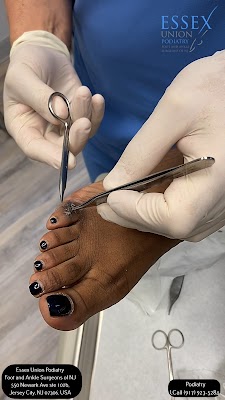
A thorough physical exam by a foot and ankle clinic surgeon guides what to image and how. Expect to stand, walk, and perform simple maneuvers. I watch how your arch behaves during a heel raise, measure ankle dorsiflexion with the knee straight and bent, and palpate for localized tenderness along tendons and joint lines. Resisted testing picks up subtle tendon tears or nerve irritability. A Tinel sign behind the medial malleolus can point to tarsal tunnel issues. Ligament testing in plantarflexion and dorsiflexion, plus peroneal tendon subluxation maneuvers, refine the problem list before a single picture is taken.
Why insist on this sequence? Imaging follows physics, not symptoms. It shows anatomy, often very well, but cannot tell me if your spring ligament laxity actually causes your medial foot pain on hills. The foot and ankle joint surgeon makes that call by putting findings into motion.
X‑rays, still the workhorse
Despite advances, standard radiographs carry a lot of weight in decision‑making. A foot and ankle bone surgeon uses them to read alignment, joint space, and bone integrity, often with precise angles. Three views of the foot and at least two of the ankle are typical. For many conditions, weight‑bearing images are essential. A flatfoot looks mild lying down and severe when the arch is loaded. A bunion’s intermetatarsal angle can expand several degrees with your full body weight. Prepare by bringing the shoes and insoles you use daily, so the foot and ankle care surgeon can match findings to how you live and work.
Special X‑ray views add detail: calcaneal axial views for heel spurs, sesamoid axial views for turf toe, and stress views for chronic ankle instability. I often obtain a hindfoot alignment view to quantify varus or valgus. Tell the tech if your pain is severe when weight bearing. There are safe ways to support you during necessary weight‑bearing shots.
Radiation dose for foot and ankle plain films is low, much less than a CT scan, and not typically a barrier. If you have had surgery with hardware, bring prior films; they guide whether a foot and ankle hardware removal surgeon needs additional Jersey City foot and ankle surgeon focused views.
Ultrasound, the dynamic storyteller
Musculoskeletal ultrasound shines in tendon and ligament evaluation, especially when performed by a foot and ankle ultrasound guided surgeon who understands both scanning and operative anatomy. It is fast, comfortable, and repeatable. Ultrasound can show fluid in tendon sheaths, partial tears, and real‑time subluxation. It also lets a foot and ankle soft tissue surgeon observe structures as you move, answering questions static imaging cannot.
Preparation is simple: wear clothing that lets the team access your ankle and midfoot. Avoid heavy lotions the day of the test. For diagnostic injections, I explain that the gel is cool and the needle is quick. If you faint with needles, tell us; we adjust positioning and monitoring.
Ultrasound has limits in deep joint cartilage or marrow edema, where MRI or CT is superior. For a Morton neuroma, though, a skilled scan can be more sensitive than MRI and can guide a small test injection that settles debates in minutes.
MRI, detail with context
MRI is the go‑to for cartilage, marrow, complex tendon pathology, and subtle stress injuries. It is invaluable for osteochondral lesions of the talus, sinus tarsi syndrome, posterior tibial tendon tears, and occult fractures. A foot and ankle advanced surgery specialist interprets MRI findings in light of the physical exam to avoid overcalling benign changes. Tendons of middle‑aged recreational athletes often show signal changes without functional deficit. Plantar fascia thickening can persist long after pain resolves.
Preparation matters more than most patients realize:
- If your pain is worse with standing, ask whether a weight‑bearing MRI is available and truly useful for your case. Not all centers have it, and not all problems benefit from it. Remove metal and tell the team if you have implants. Many foot screws and plates are MRI‑compatible, but older pacemakers or certain neurostimulators may preclude MRI. If you are claustrophobic, request an open unit or mild anxiolytic in advance. Appointments run 20 to 45 minutes, depending on sequences.
Gadolinium contrast is rarely needed for routine tendon or ligament imaging but becomes important for infection workups or postoperative scar versus retear. If renal function is impaired, your foot and ankle infection surgery specialist will balance contrast benefits against risks and, in some cases, favor nuclear medicine or CT.
CT and weight‑bearing CT, the blueprint of bone
CT excels at bony detail, union assessment, malunion mapping, and preoperative planning for osteotomies and fusions. A foot and ankle osteotomy surgeon studies three‑dimensional deformity that plain films can hide, including calcaneal tuberosity orientation, subtalar coalition, and talonavicular congruency. Surgical planning software linked to CT data can help a foot and ankle custom surgical plan doctor simulate cuts and implant positions that match your anatomy.
Weight‑bearing CT, available in more centers now, changes the game for flatfoot collapse, cavus alignment, and midfoot arthritis. It shows how your joints load in real life. The dose is higher than plain films, lower than full conventional CT, and often justified when alignment drives your pain.
Prepare by bringing prior imaging on disc. If the clinic uses digital transfer, confirm the files include the full DICOM set, not screenshots. Positioning matters; I check whether you stood in your neutral stance or were rotated.
CT arthrogram, where contrast is injected into a joint before CT, is sometimes preferred over MRI for cartilage flap detection in the ankle. It requires a radiologist or foot and ankle procedure specialist comfortable with sterile injections. If you have an iodine contrast allergy, premedication may be needed or an alternate test chosen.
Nuclear medicine, bone scans, and SPECT‑CT
These studies come into play for infection, complex post‑operative pain, suspected nonunion, and cases where MRI cannot be performed. A combined SPECT‑CT overlays metabolic activity on anatomy, helping a foot and ankle non union repair surgeon identify a symptomatic joint in a multifocal arthritic foot. Preparation includes verifying medications, allergies, and pregnancy status. The test involves tracer injection and imaging over several hours.
Electrophysiology and vascular testing
Nerve conduction studies and EMG help when tingling, burning, or numbness complicate the picture. A foot and ankle nerve entrapment surgeon looks for tibial nerve compression at the tarsal tunnel, superficial peroneal nerve neuritis at the fibular neck, or radiculopathy from the back. The test is uncomfortable but brief. Clinical correlation is key, since false positives and negatives exist.
For suspected vascular compromise, especially in smokers, diabetics, or older patients, ankle‑brachial indices and Doppler ultrasound guide wound and fusion risk. A foot and ankle geriatric surgery specialist may enlist a vascular colleague if pulses are weak or wounds are slow to heal.
Diagnostic injections: the fastest truth serum we have
Targeted local anesthetic injections separate overlapping pain sources. If numbing the sinus tarsi erases your lateral hindfoot pain during a hallway walk, subtalar impingement moves up the list. If an intra‑articular ankle injection provides full relief, cartilage or osteophytes likely drive symptoms more than peroneal tendinopathy. A foot and ankle ultrasound guided surgeon maximizes accuracy and minimizes steroid dose when steroids are used.
For gout versus infection in a hot, swollen first MTP joint, aspiration answers what pictures sometimes cannot. Crystals under a microscope can settle the question in minutes. The foot and ankle gout surgery doctor still cares about tophi and joint damage visible on imaging, but the presence of monosodium urate changes your whole treatment path.
Labs that matter more than length
Ordered sparingly and strategically, labs support clinical judgment. ESR, CRP, and CBC inform infection risk. Uric acid supports gout but does not diagnose it alone. Rheumatologic panels enter when multiple joints flare or deformities progress quickly, guiding a foot and ankle rheumatoid surgery specialist toward joint preservation strategies in collaboration with rheumatology. Vitamin D and calcium data can matter in nonunion risk and bone graft planning.
Building the plan: how tests influence candidacy and technique
A foot and ankle surgical evaluation doctor uses a structured approach to decide who benefits from surgery and what operation gives the best odds of durable relief. Imaging and tests feed several core decisions:

- Is the primary lesion consistent with symptoms, and has it failed reasonable conservative care for a sufficient trial, often 6 to 12 weeks? Are biomechanics correctable by orthoses or bracing, or does deformity require realignment by a foot and ankle alignment correction surgeon? What is tissue quality? Tendons with diffuse degeneration may not hold sutures well, steering the foot and ankle structural repair surgeon toward augmentation or transfer rather than simple repair. How stable are neighboring joints? A medial column collapse often pairs with equinus; missing either invites failure. A foot and ankle gait correction surgeon looks at the chain, not just the link. What risks change the calculus: diabetes control, smoking, neuropathy, vascular disease, or immunosuppression? A foot and ankle surgical risk assessment specialist addresses these first.
When tests argue for surgery, they also refine the method. An osteochondral lesion contained and smaller than roughly 1 to 1.5 cm may suit microfracture. Larger or cystic lesions may benefit from autograft, allograft, or particulated cartilage working with a foot and ankle cartilage repair surgeon. A subtle syndesmotic injury on MRI, if unstable on stress radiographs, may push the foot and ankle internal fixation surgeon to dynamic stabilization with a suture button rather than rigid screws for certain athletes.
What to bring and how to prepare for your imaging visit
- All prior imaging on disc or secure link, plus reports. Include studies from the knee and back if nerve symptoms exist. A short treatment timeline with dates, devices, therapy notes, and responses. Pictures of swelling or shoe wear patterns help. Shoes and orthotics you wear most, and the pair that hurts you most. A comprehensive medication and allergy list, including reactions to iodine or gadolinium. Questions written down, prioritized by what you need answered before you leave.
Day‑of testing details that prevent delays
Small steps prevent rescheduling. Eat normally unless told otherwise. If contrast is planned and you have diabetes on metformin, ask if you should hold it. Remove ankle bracelets and toe rings. Wear loose pants that can roll above the calf. Tell the technologist where it hurts most and when. Weight‑bearing images should mimic your true stance, not a forced posture.
For MRI, arrive early to complete safety forms. If you have a known contrast allergy, call the office two to three days before to arrange premedication if contrast cannot be avoided. If you have a pacemaker or spinal cord stimulator, bring the device card. For ultrasound‑guided injections, arrange a ride if a numbed joint will make driving unsafe.
Radiation is a common worry. A four‑view ankle series delivers a tiny fraction of the dose of a CT. A foot CT without contrast typically falls far below a chest CT. We avoid repeat studies by coordinating with your imaging center and verifying image quality before you leave.
Reading the pictures without letting them read you
Patients often arrive with bold MRI phrases. “High‑grade partial tear.” “Severe tendinosis.” “Multifocal bone marrow edema.” Those words carry weight, but context rules. A foot and ankle operative specialist compares those findings to your specific pain and function. For example:
- Many asymptomatic adults show plantar fascia thickening. If your pain is lateral and worsens with eversion, peroneal overload is more likely than recalcitrant fasciitis. Bone marrow edema around the ankle is common after a twist, sometimes persisting for months. On its own it rarely justifies surgery. Tiny longitudinal tendon splits can be normal in chronic mileage runners. True surgical targets tend to produce reproducible weakness, subluxation, or focal, load‑dependent pain.
I prefer to review your images with you. We look at the same slice that the report mentions, then we press on the matching spot on your ankle. Alignment views often lead to “aha” moments, because alignment connects directly to how your foot behaves in a shoe.
When repeating or expanding tests is wise
Not every first study answers the question. Here are judgment calls that pay off:
- If the only MRI is non weight‑bearing and your symptoms scream mechanical collapse, add weight‑bearing X‑rays or a weight‑bearing CT. If the radiologist mentions a cartilage lesion but your pain is vague, consider a diagnostic intra‑articular injection before committing to a cartilage procedure. If nerve symptoms change, repeat EMG/NCS after 8 to 12 weeks to track recovery or progression. If infection is suspected but MRI is blurred by metal, use tagged white blood cell scans or SPECT‑CT.
The foot and ankle surgical second opinion process often centers on this kind of tailoring rather than simply re‑reading the same report.
Special considerations: kids, older adults, and athletes
Children are not small adults. Growth plates mimic fractures on imaging. A foot and ankle growth plate surgeon reads these carefully, correlating with tenderness and function. Sedation for MRI is sometimes required; this is planned with pediatric anesthesia. Avoiding unnecessary radiation is a priority, so ultrasound and MRI are favored when possible. For flexible flatfoot with pain, weight‑bearing films and hindfoot alignment views matter; for rigid flatfoot, consider coalition with CT.
Older adults bring different flags: bone density, vascular status, and balance. A foot and ankle geriatric surgery specialist may order a DEXA scan in cases of nonunion risk or fragility fractures. Doppler assessment can change wound planning, and skin quality dictates incision choices for a foot and ankle minimally scarring surgeon.
Athletes need load‑specific data. A ballet dancer’s posterior impingement appears on plantarflexion stress views, not just neutral films. A soccer player’s syndesmotic sprain may hide until external rotation stress is applied. Return‑to‑play timelines depend on both pictures and performance metrics. A foot and ankle accelerated recovery surgeon blends biologic healing data with strength and balance testing.
Two brief vignettes from clinic
A weekend hiker with chronic lateral ankle pain arrived with a normal non weight‑bearing MRI. Exam showed tenderness over the sinus tarsi and a positive single‑leg balance test. Weight‑bearing CT revealed lateral impingement between calcaneus and fibula during stance. A diagnostic injection into the sinus tarsi relieved pain immediately. The final plan mixed orthotic posting and a small arthroscopic debridement by a foot and ankle endoscopic surgery specialist. Without the weight‑bearing study and targeted injection, she was heading toward an unnecessary peroneal repair.

A chef with a two‑month‑old ankle fracture repair could not shake deep ache. X‑rays looked acceptable. CT, however, showed a small posterior malreduction that loaded the posterior tibial plafond. Vitamin D was low, and he smoked ten cigarettes a day. A foot and ankle post surgical revision specialist staged care: vitamin D repletion, smoking cessation, and a bone stimulator. Pain improved, but not enough. After three months and consistent labs, a foot and ankle advanced reconstruction doctor performed a limited revision that restored posterior alignment. He returned to full shifts without limp. The key was targeted CT rather than guesswork.
Questions to ask your foot and ankle surgical evaluation doctor
- Which imaging or tests will change the plan, and which are routine? If we skip one, what do we risk missing? For my pain pattern, should images be weight bearing, and do we need stress views or dynamic ultrasound? If surgery is on the table, what measurements from these tests decide the technique, implants, or need for bone graft? Are there diagnostic injections we can try to confirm the source before I commit to an operation? How will my medical conditions, medications, or implants affect imaging choices, anesthesia, or healing?
Cost, access, and practical timing
Not everyone has immediate access to MRI or weight‑bearing CT. Insurance often requires a sequence: X‑ray, conservative care, then advanced imaging. A foot and ankle surgical referral specialist can document exam findings and functional limits in a way that speeds approval when advanced imaging is clearly needed. If delays stretch, targeted ultrasound and diagnostic injections can maintain progress. Ask the team whether same‑day imaging is available. In many clinics, a foot and ankle outpatient surgeon can perform ultrasound‑guided injections during the first visit if the diagnosis is clear enough.
For those paying out of pocket, prices vary widely. Plain films might range from tens to low hundreds of dollars, MRI from several hundred to over a thousand depending on setting, and CT somewhere in between. Self‑pay programs at imaging centers can cut list prices significantly. If a test will not change your plan, do not order it. A foot and ankle evidence based surgeon should say that plainly.
After the tests: translating data into a plan you can live with
The end of a good evaluation is not just a stack of images but a path. Sometimes that path avoids the operating room. Bracing, targeted therapy, shoe modifications, and ultrasound‑guided tendon procedures like PRP, if indicated, can settle many cases. A foot and ankle regenerative surgery specialist will help you understand when biologic injections might add value and when they are unlikely to help. If surgery makes sense, the data shape a custom plan: incision placement guided by ultrasound findings, implant choice guided by bone quality on CT, and alignment targets based on weight‑bearing angles.
A foot and ankle joint preservation surgeon may favor osteotomy and soft‑tissue balancing in young patients with isolated deformity. A foot and ankle arthritic joint surgeon might counsel fusion over replacement in heavy laborers or severe deformity. A foot and ankle robotic assisted surgeon, where available, can use CT‑based planning to improve accuracy in ankle arthroplasty. Every technique has trade‑offs. A frank discussion of risks, graft needs, and expected recovery timelines belongs in the same room as your images, not in a handout.
Follow‑through matters. Postoperative radiographs confirm hardware and alignment. Ultrasound can track tendon glide after repair. A foot and ankle surgical recovery specialist uses both pictures and milestones, like pain‑free single‑leg stance and a symmetric heel raise, to pace your return. If complications arise, prompt imaging by a foot and ankle complication management surgeon can catch a brewing nonunion or infection before it derails your outcome.
The quiet power of preparation
Imaging does not heal a tendon or straighten a bone, but it can prevent wrong turns. When you show up with a precise story, prior studies organized, and a few targeted questions, you help your foot and ankle medical surgeon focus the right tests on the right target. In turn, a thoughtful foot and ankle surgical consultant will choose the smallest set of studies that yield the largest clarity, interpret them in motion, and build a plan that fits your life.
That runner with the scary MRI? Dynamic ultrasound showed a stable peroneal tendon with mild sheath swelling. A weight‑bearing X‑ray revealed a subtle cavus foot and a plantarflexed first ray. A small orthotic post, calf stretching, and a single ultrasound‑guided injection calmed the pain. No knife, just the right tests read the right way.
If surgery is ahead, or if your goal is to avoid it, preparation for imaging and tests is not busywork. It is the backbone of clear diagnosis and smart treatment by the foot and ankle surgical team you are trusting with every step.